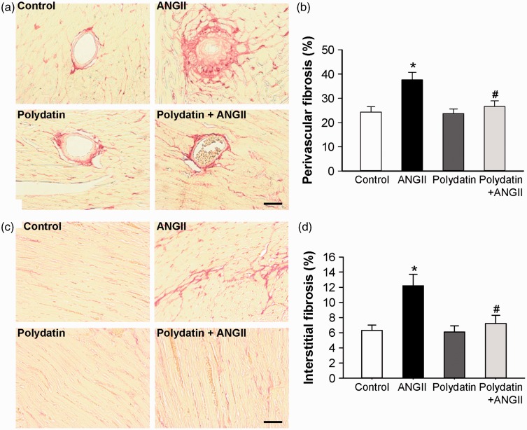
Figure 4.
Pretreatment with polydatin inhibits left ventricles fibrosis induced by Ang II in rats. Left ventricles fibrosis was measured in the rats treated with control vehicle, Ang II infusion, polydatin, or Ang II infusion plus polydatin. (a) Representative images of intramuscular arteries with perivascular fibrosis stained with picric-sirius red. (b) Bar graph shows quantified perivascular fibrotic areas (%). (c) Representative images of myocardium with interstitial fibrosis stained with picric-sirius red. (d) Bar graph shows quantified interstitial fibrotic areas (%). The scale in the images is 50 µm. Results were expressed as means ± SE (n = 6). *P < 0.05 versus control values. #P < 0.05 versus Ang II alone. (A color version of this figure is available in the online journal.)

