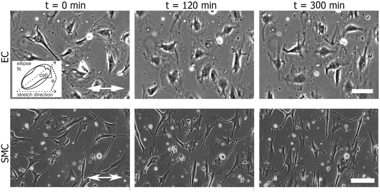
Figure 1.
The alignment of vascular cells with respect to the direction of a uniaxial cyclic tensile strain application. Human primary endothelial cells (ECs) and smooth muscle cells (SMCs) adherent on fibronectin-coated silicone surfaces were exposed to uniaxial cyclic tensile strain. Still images of ECs and SMCs from phase contrast movies show the cells before (t = 0 min) and after 120 and 300 min of uniaxial cyclic strain with a frequency of 1 Hz. The direction of strain is indicated by the white double-headed arrow. The scale bar represents 200 µm. Inset: Scheme of ellipse fitting for analysis of cell orientation; the angle ϕ is between the ellipse main axis and the stretch direction

