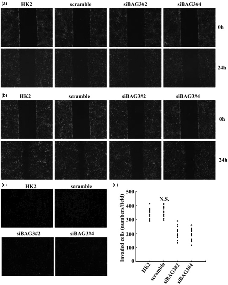
Figure 4.
Suppression of FGF-2-mediated motion and invasion of HK2 cells by BAG3 knockdown. (a) HK2 cells were transduced with siBAG3 or scramble lentiviral vectors for 12 h and cultured for additional 48 h. Cell motility was analyzed using wound-healing assays. (b) HK2 cells were transduced with siBAG3 or scramble lentiviral vectors for 12 h and cultured for additional 48 h. Cells were then treated with 10 ng/mL of FGF-2 and cell motion was evaluated using wound-healing assays. (c) HK2 cells were transduced with siBAG3 or scramble lentiviral vectors for 12 h and cultured for additional 48 h. Cells were then treated with 10 ng/mL of FGF-2 and cell invasion was evaluated by Matrigel-coated Transwell system. (d) Cells that have passed through Matrigel-coated membrane in (c) were counted in five representative microscopic fields and three independent experiments were performed. Cell numbers for each count were plotted in the graph. N.S.: not significant; HK2: human kidney 2. *P < .01. (A color version of this figure is available in the online journal.)

