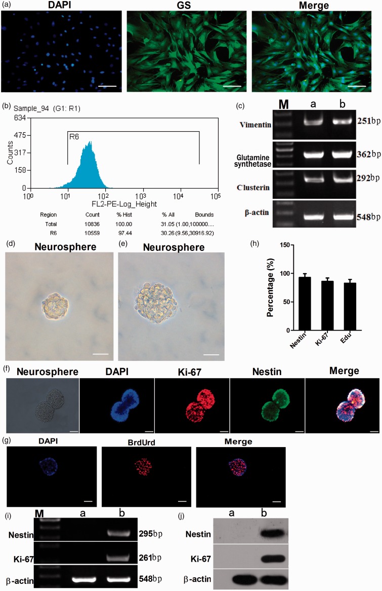
Figure 1.
Characterization of enriched Müller cells. Immunocytochemical analysis showed that more than 98.10% of retinal Müller cells were immunoreactive to Müller cell marker GS (a). Bar = 200 µm. FACS showed that 97.44% of purified cells were immunoreactive for GS (b). The purity of enriched cells was evaluated by RT-PCR analysis to detect the expression of Müller cell-specific transcripts (c). Enriched Müller cells exposed to the stem cell-conditioned medium formed neurospheres (d). Bar = 200 µm. The neurospheres were passaged to get new clonal neurospheres (e). Bar = 200 µm. Immunofluorescence staining showed that the stem cells within the cell spheres had positive expression of retinal stem cell-specific markers Nestin (92.94 ± 6.48%) and Ki-67 (85.96 ± 6.04%) (f, h). Bar = 100 µm. Immunocytochemical analysis of BrdUrd showed that newborn cell spheres had the capacity of effective proliferation (82.80 ± 6.65%) (g, h). Bar = 100 µm. RT-PCR and Western blot analysis to detect the expression of stem cell markers Nestin and Ki-67 (i, h). c: Lane M: DNA marker; lane a: PN21 retina; lane b: purified Müller cells. i,j: lane M: DNA marker; lane a: Müller cells; lane b: neurospheres. (A color version of this figure is available in the online journal.)

