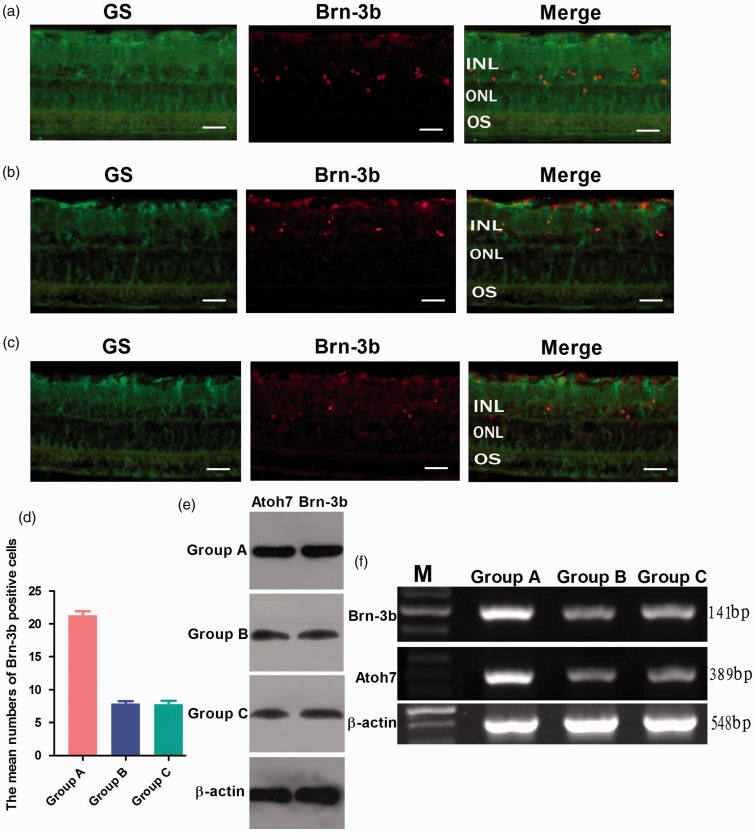
Figure 4.
Differentiation of retinal stem cells in rat chronic ocular hypertension glaucoma model. Immunocytochemical staining of retinal tissue sections showed that the proportion of Brn-3b positive cells was 21.3 ± 2.0% in group A, but only 7.9 ± 1.1 and 7.8 ± 1.7% of positive cells expressed Brn-3b in group B and C (a–d). Western blot and RT-PCR analysis showed that the expression of Atoh7 and Brn-3b at both mRNA and protein levels was significantly increased in group A. There were no significant differences between group B and group C (e, f). e,f: lane M: DNA marker; group A: transfected with lentivirus PGC-FU-Atoh7-GFP; group B: transfected with empty vector PGC-FU-GFP; group C: no transfection. Bar = 100 µm. (A color version of this figure is available in the online journal.)

