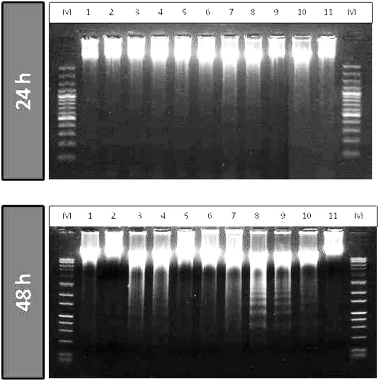
Figure 5.
DNA fragmentation in HepG2 cells after 24 h and 48 h of treatment with long chain fatty acid esters of Q3G. Cells (5 × 10⁵ cells; 12-well plate) were treated with 100 µM of the test compounds for 24 h and 48 h. Cells were collected, lysed and DNA was extracted and run on agarose gel containing GelRed™ DNA staining solution for fragmentation analysis. Lane M, DNA marker; lane 1, Cisplatin; lane 2, stearic acid ester of Q3G; lane 3, oleic acid ester of Q3G; lane 4, linoleic acid ester of Q3G; Lane 5, alpha-linolenic acid ester of Q3G; lane 6, EPA ester of Q3G; lane 7, DHA ester of Q3G; lane 8, Q3G; lane 9, quercetin; lane 10, Sorafenib; lane 11, control (no treatment). After 48 h of treatment all long chain fatty acid esters of Q3G except stearic acid ester treatment showed substantial amount of apoptosis as seen by the fragmented DNA pattern

