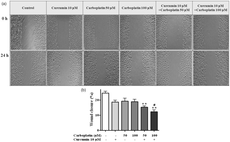
Figure 3.
Wound healing assay. After the cells had formed a confluent monolayer, scratches in the monolayer were made using a 200 µL tip and closure of the scratch was examined under a microscope. (a) Representative photographs show the same area at 0 h and after 24 h of treatment. (b) Cells migrating into the wound area were counted based on the line of the wound at 0 h. *, P < 0.05; **, P < 0.01 compared with control; #, P < 0.05 compared with 100 μM carboplatin

