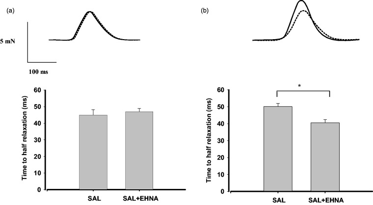
Figure 4.
β2 adrenoceptor-mediated lusitropic responses to salbutamol (SAL) in right and left ventricular myocardium. Strips of rat right ventricle (a) or left ventricular papillary muscle (b) were exposed to 3 µmol/L salbutamol plus 300 nmol/L CGP-20712A to block β1 adrenoceptors either in the absence (dotted lines) or in the presence (solid lines) of the PDE2 inhibitor EHNA (10 µmol/L). Top: original registration. Bottom: time required to half relaxation time. Data are expressed as means ± S.E.M. of six experiments. *P < 0.05

