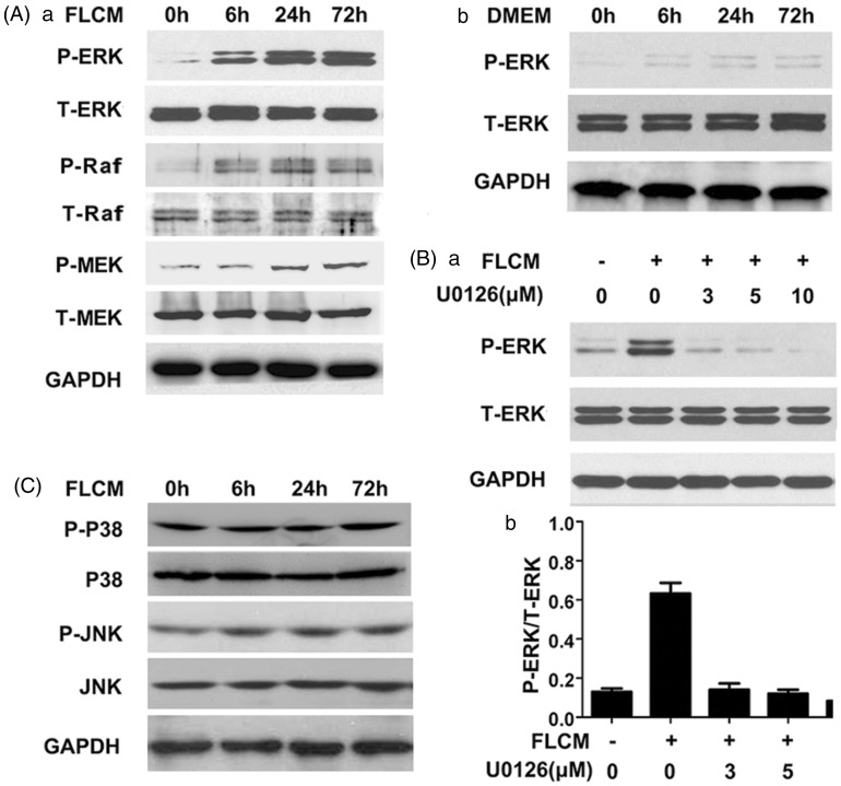
Figure 5.
FLCM provoked ERK1/2 phosphorylation in hUCMSCs. A: Western blotting analysis of P-ERK, P-Raf, P-MEK, and T-ERK, T-Raf, T-MEK in FLCM-treated (a) and DMEM-treated (b) hUCMSCs at 0, 6, 24, and 72 h; B: Effect of U0126(ERK1/2 inhibitor) on the ERK1/2 phosphorylation at 3, 5, and 10 µM after FLCM treatment for 24 h (a). Density analysis of Western blotting bands (b). Data are expressed as mean + SD of three experiments. C: Western blotting analysis of P-JNK, JNK, P-P38, and P38 in FLCM-treated hUCMSCs at 0, 6, 24, and 72 h. P-ERK, phosphorylated ERK1/2; T-ERK, total ERK 1/2; P-Raf, phosphorylated Raf; T-Raf, total Raf; P-MEK, phosphorylated MEK; T-MEK, total MEK

