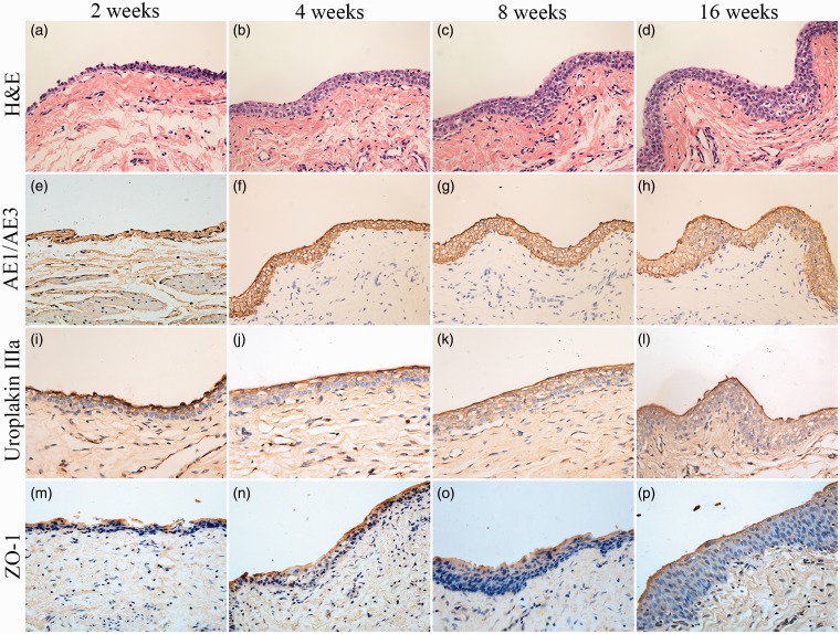
Figure 6.
Histologic characteristics of TETSs in the experimental group after urinary diversion at 2, 4, 8, and 16 weeks. (a, b, c, and d) H&E staining displayed the regeneration of epithelium layers of TETSs (× 400). (e–h, i–l, and m–p) Immunohistochemical staining of AE1/AE3, uroplakin IIIa, and ZO-1 revealed the regeneration of epithelium of TETSs, respectively (× 400). (A color version of this figure is available in the online journal.)

