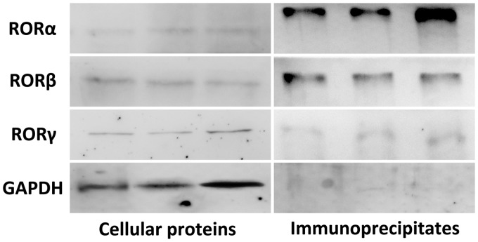
Figure 5.
The interaction between SERPINA3K and RORs. Proteins were extracted from normally cultured MS1 cells. Western blot analysis used the whole cellular protein was conducted to verify the expression of RORs (left panel). Anti-SERPINA3K polyclonal antibody was added into the cellular proteins to produce protein-antibody immunoprecipitates, which were collected by protein A/G PLUS-Agarose. Western blot was applied using this protein-anti-SERPINA3K immunoprecipitates, and found apparent straps of RORα and RORβ, while the strap of RORγ was hardly to see (right panel)

