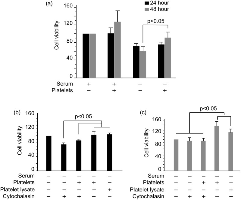
Figure 3.
Platelets promote HUVEC viability. (a) HUVECs were plated at 5 × l04 cells/mL in 96-well plates in ECM supplemented with 10% FBS and incubated for 24 h. Next, the medium was replaced with serum-deprived or 10% FBS-containing ECM and platelets were added. Cells were cultured continuously for 24 and 48 h. Cell viability was determined using the MTT assay. Error bars represent the SD value. (b, c) HUVECs were cultured in serum-deprived ECM in combination with platelets, cytochalasin B, or platelet lysates for 24 (b) and 48 h (c). Cell viability was determined using the MTT assay. Data are expressed as the mean and SD values of quadruplicate wells of three independent experiments

