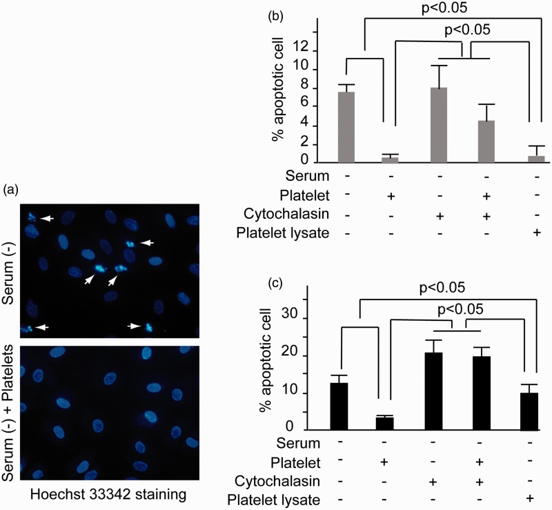
Figure 4.
Inhibition of HUVEC apoptosis by platelet phagocytosis. (a) HUVECs were cultured in the serum-deprived ECM in the presence or without platelet for 48 h. Then Hoechst33342 staining analysis was performed. The dense nuclear condensation and shrinkage marked by arrows indicated apoptotic cells. Magnification × 400. (b) HUVECs were cultured in serum-deprived ECM together with platelets, platelet lysate, or cytochalasin B, respectively, for 48 h. Then the apoptotic ratio of HUVECs was examined by Hoechst33342 staining analysis. (c) HUVECs were cultured in serum-deprived ECM in the presence of platelets, platelet lysate, or cytochalasin B, respectively, for 72 h. Then the apoptotic ratio of HUVECs was examined by Hoechst33342 staining analysis. Significant inhibition of apoptosis was noted in the platelet and platelet lysate incubation groups (p < 0.05). All of the experiments were performed thrice. (A color version of this figure is available in the online journal.)

