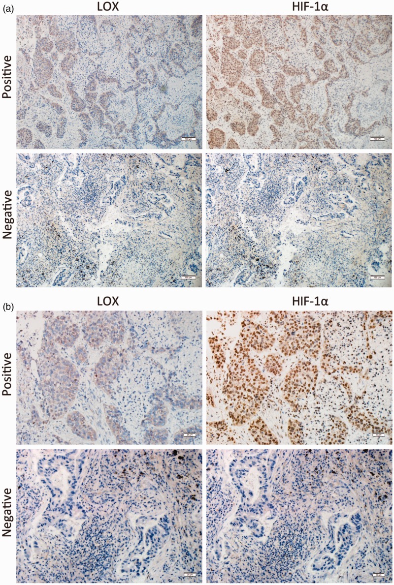
Figure 1.
LOX was correlated with HIF-1α expression in patients with NSCLC. Continuous section of NSCLC was obtained and subjected to anti-LOX and anti-HIF-1α antibodies. (a) Representative micrographs for immunohistochemical staining of HIF-1α (right) and LOX (left) were shown (Magnification: 100×). (b) Details of immunohistochemical staining were shown (Magnification: 400×). (A color version of this figure is available in the online journal.)

