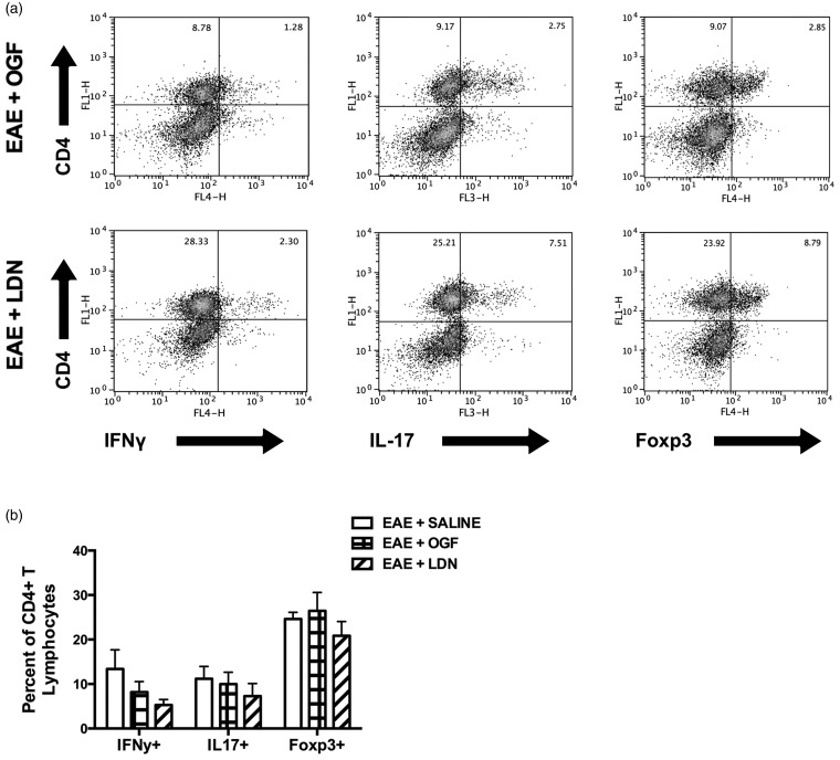
Figure 3.
Effector CD4+ T lymphocytes isolated from the brain and spinal cord of EAE mice after five days of treatment (10 mg/kg OGF, 0.1 mg/kg naltrexone (LDN), or saline), corresponding to 15 days post immunization. (a) Intracellular cytokine staining of IFNγ, IL-17, and Foxp3 in mononuclear cells isolated from CNS tissue gated on live cells after ex vivo stimulation with PMA/ionomycin and brefeldin A for 4 h. (b) Percentage of CD4+ T lymphocytes that are positive for IFNγ, IL-17, and Foxp3 are shown.
EAE: experimental autoimmune encephalomyelitis; OGF: opioid growth factor; LDN: low dosage of naltrexone

