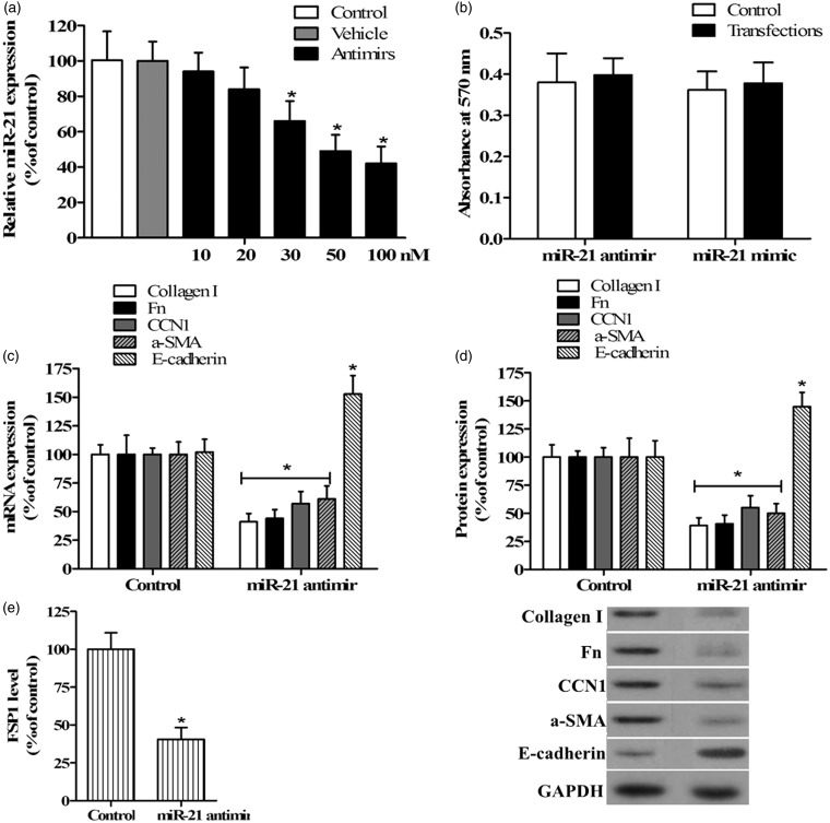
Figure 2.
Inhibition of miR-21 affected ECM protein and EMT marker molecule expression in TGF-β-stimulated TECs. (a) qRT-PCR was used to analyze the expression of miR-21 in TECs transfected with 10, 20, 30, 50, and 100 nM miR-21 antimir and 100 nM control miRNA (vehicle). (b) The MTT method was used to determine the viability of cells transfected with miR-21 antimir or mimic, and absorbance was measured at 570 nm. Untransfected cells were used as the control in a and b. qRT-PCR (c) and Western blotting (d) were used to analyze the expression of type I collagen, Fn, CCN1, α-SMA, and E-cadherin in TECs stimulated with TGF-β after transfection with miR-21 antimir or control miRNA, as shown in representative Western blots. FSP1 level was detected by ELISA (e). Cells transfected with control miRNA were used as the control in c, d, and e (n = 5/group, *P < 0.05 versus control)

