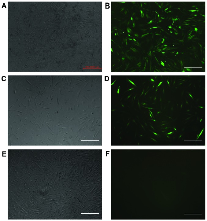
Figure 2.
The cells were observed under a fluorescence microscope (×50 magnification) on the 4th day post-transfection. (A and B) Cells were positive for the expression of green fluorescent protein (GFP) in the integrin-linked kinase (ILK)-siRNA-LV-transfected group; (C and D) GFP expression was also positive in the negative control LV-transfected group. (E and F) GFP expression was negative in the normal control group. Scale bars, 500 µm.

