. 2016 Jun 2;5:e14226. doi: 10.7554/eLife.14226

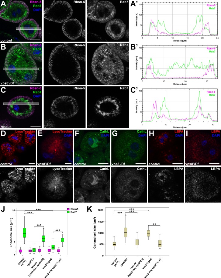
Figure 5. The biogenesis of late endosomes and lysosomes is impaired in vps8 mutant garland nephrocytes.

(A–C) Late endosomes are fragmented in garland nephrocytes lacking Vps8, unlike early endosomes. In wild type control or rescued cells, a layer of Rab7-positive endosomes (green) is found under the peripheral Rbsn-5 positive endosomes (magenta). In contrast, small granular Rab7 structures fill the whole cytoplasm of vps8 mutants, while the Rbsn-5 pattern remains unchanged in these cells. Panels (A’–C’) show the plot profiles of the framed areas in composite images. (D,E) LysoTracker-, (F,G) Cathepsin L- (H,I) and LBPA-positive lysosomal structures are fragmented in vps8 mutant nephrocytes. (J) Quantification of data from panels (A–C) and from Figure 5—figure supplement 1. (K) Vps8 mutant nephrocytes are significantly larger than control or rescued cells. The medians of data are indicated as horizontal black lines within the boxes. Bars show the upper and lower quartiles, **p<0.01, ***p<0.001. Bars: 10 µm. Please see Figure 5—figure supplements 1,2 for additional data.

DOI: http://dx.doi.org/10.7554/eLife.14226.010

Figure 5.

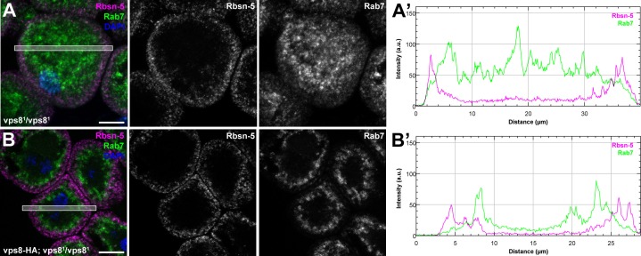
Figure 5—figure supplement 1. The biogenesis of late endosomes is impaired in homozygous vps8 mutant garland nephrocytes.

Figure 5—figure supplement 1.

(A) Rab7 positive late endosomes are fragmented in homozygous vps81 mutant garland nephrocytes, similar to vps81/Df cells shown in Figure 5B. (B) Late endosome defects of vps81 homozygotes are rescued by the vps8-HA transgene. Bars: 10 µm. Panels (A’) and (B’) show the plot profiles of the framed areas in composite images.
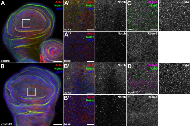
Figure 5—figure supplement 2. The endosomal compartment of larval wing disc cells is normal in vps8 mutant larvae.

Figure 5—figure supplement 2.

(A,B) Larval wing discs stained with cortical actin phalloidin and extracellular domain-specific anti-Notch. Wing disc structure and the pattern of endosomal Notch are similar in control (A) and vps8 mutant (B) animals. Panels (A’, B’) and (A’’, B’’) show high magnification images of the framed areas in (A) and (B), respectively, and are taken from the apical and basal regions of the columnar cell layer. (C,D) High magnification images showing the apical regions of the columnar cell layer at the wing pouch region of the discs, stained with anti-Rbsn-5 and anti-Rab7. The pattern and distribution of both early and late endosomes are similar in controls (C) and vps8 mutants (D). Bars: (A, B): 50 µm, (A’–B’’), (C,D): 10 µm.