Fig. 4.
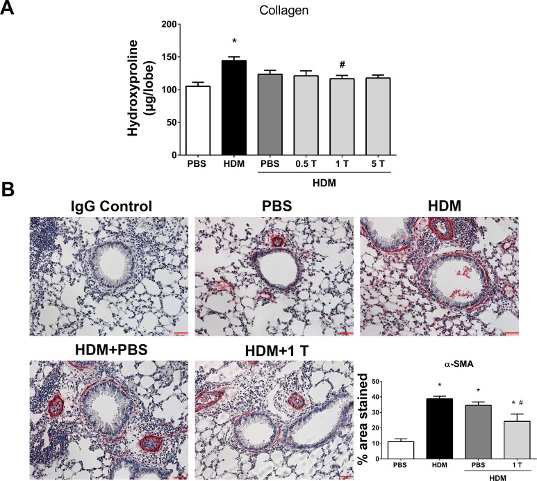
Preventive regimen of TUDCA decreases HDM-induced airway fibrosis. A: measurement of collagen by hydroxyproline assay. Data are means ± SE of 6–8 mice/group. *P < 0.05 compared with their respective PBS controls and #P < 0.05 compared with vehicle-untreated HDM-challenged mice. B: representative images of α-smooth muscle actin (α-SMA)-stained lung tissue sections of vehicle-treated or untreated and TUDCA (1 mg/kg body wt dose)-treated HDM-challenged mice (×20 magnification; scale bars = 50 μm) and the quantification of percentage of area that positively stained for α-SMA immunostaining. Data are means ± SE of 6–8 mice/group. *P < 0.05 compared with their respective PBS controls and #P < 0.05 compared with vehicle-untreated HDM-challenged mice.

