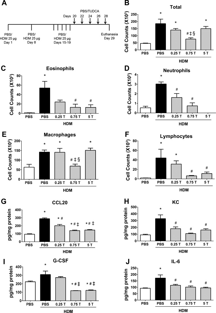
Fig. 5.
Therapeutic regimen of TUDCA decreases HDM-induced airway infiltration of inflammatory cells and decreases inflammatory cytokines and chemokines. A: schematic representing the time points of HDM or PBS instillation and TUDCA treatment. HDM (25 μg/mouse) was instilled intranasally while TUDCA (0.25, 0.75, and 5 mg/kg body wt) was administered via nasopharynx after the HDM challenge phase. B–F: analysis of inflammatory and immune cells in the BALF. Data are means ± SE of 6–8 mice/group. *P < 0.05 compared with their respective PBS controls. #P < 0.05 compared with vehicle-treated HDM-challenged mice. ‡P < 0.05 compared with mice treated with 0.25 mg/kg TUDCA. §P < 0.05 compared with mice treated with 5 mg/kg TUDCA. G–J: ELISA for cytokines and chemokines. Data are means ± SE of 4–6 mice/group. *P < 0.05 compared with their respective PBS controls. #P < 0.05 compared with vehicle-treated HDM-challenged mice. ‡P < 0.05 compared with mice treated with 0.25 mg/kg TUDCA.

