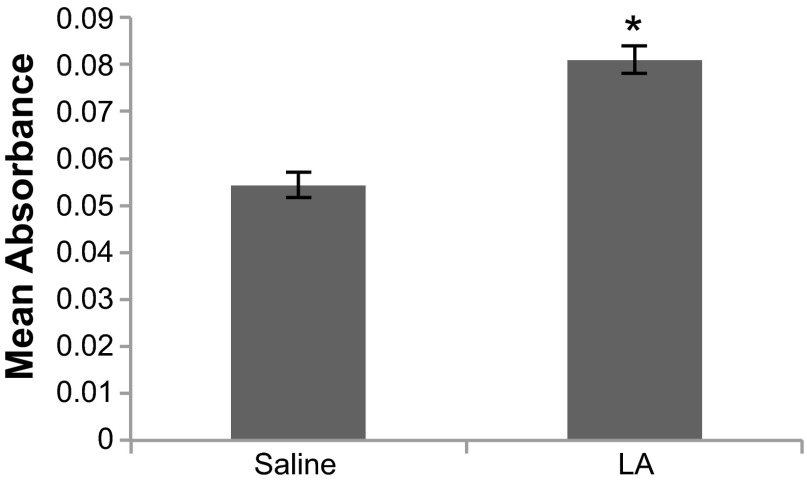
Fig. 2.
Binding ELISA with mouse mesothelial cells to demonstrate the presence of mesothelial cell autoantibodies (MCAA). Sera were pooled from mice exposed it to saline only or to 30 μg Libby Amphibole (LA) in sterile saline for 7 mo. Mouse primary mesothelial cells were plated in 96-well plates and then stained for MCAA binding using the pooled serum as the primary antibody and anti-mouse IgG-horseradish peroxidase (HRP) for the secondary antibody. n = 3 Wells/treatment group. *P < 0.05 by 2-tailed t-test.

