Fig. 3.
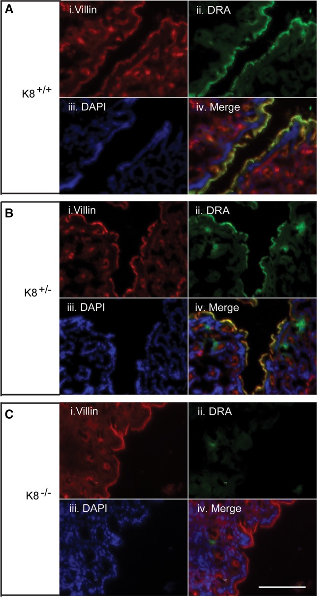
Immunofluorescence staining of distal colon shows the dramatic loss of DRA in K8−/−, but not in K8+/− mice. Distal colon cryosections fixed with 4% paraformaldehyde in PBS for 15 min at room temperature were stained with DRA. The results show absence of DRA in K8−/− mice (C) compared with normal DRA staining in K8+/+ mice (A). K8+/− mice showed patchy DRA staining (B). DRA was abolished in the K8−/− distal colon apical membrane compared with K8+/− and K8+/+ mice, whereas apical villin was normally distributed. DAPI was used to stain nuclei. Scale bar 100 μm.

