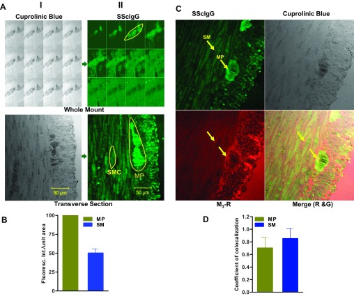
Fig. 1.
A: (i) Identification of myenteric neuronal plexus (MP) by cuprolinic blue staining on whole mount z-stack and transverse section (ii) staining of MP and smooth muscle(SM) by SScIgG; Anti human FITC-conjugated-antibody was used against SScIgG, which gives green florescence. B: bar graph of IFI showing that the binding intensity of SScIgG to MP is significantly higher compared with SM (P < 0.05; n = 3). C: immunohistochemical colocalization of SScIgG (FITC-conjugated; green) and M3-R (TR-conjugated; red). Arrows indicate colocalization of both probes on SM and MP (P < 0.05; n = 3). D: bar graph data show significant coefficient of colocalization of SScIgG with the M3-R at SM and MP (P < 0.05; n = 3).

