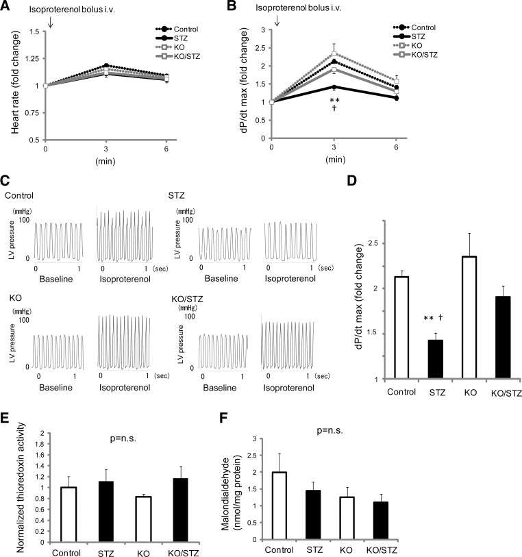
Fig. 3.
Stress in vivo hemodynamic analysis uncovers the evidence of masked mechanical dysfunction by hyperglycemia and that Txnip-KO hearts have a greater inotropic reserve under diabetic conditions. Hemodynamic parameters were acquired under anesthesia with a catheter positioned in the left ventricle (LV). Isoproterenol (1.6 ng/g BW) was given to animals by intravenous bolus injection via the right jugular vein. A: β-adrenergic challenge minimally increased heart rate at 3 min after the injection in wild-type nondiabetic (Control), streptozotocin-induced diabetic (STZ), Txnip-KO nondiabetic (KO), and Txnip-KO diabetic (KO/STZ) mice. B: the maximal rate of rise of LV pressure (dP/dt max) was increased by isoproterenol injection. C and D: representative traces of LV pressure and comparisons of dP/dt max among 4 groups at 3 min, respectively. Data are expressed as means ± SE. n = 4–9. **P < 0.01 between Control and STZ; †P < 0.05 between STZ and KO/STZ. E and F: despite these mechanical functional differences, neither thioredoxin activities (E) nor tissue levels of malondialdehyde (F), both indicators of oxidative stress, were different in whole heart homogenates among the groups. P = N.S. n = 4 or 5 each.

