Fig. 7.
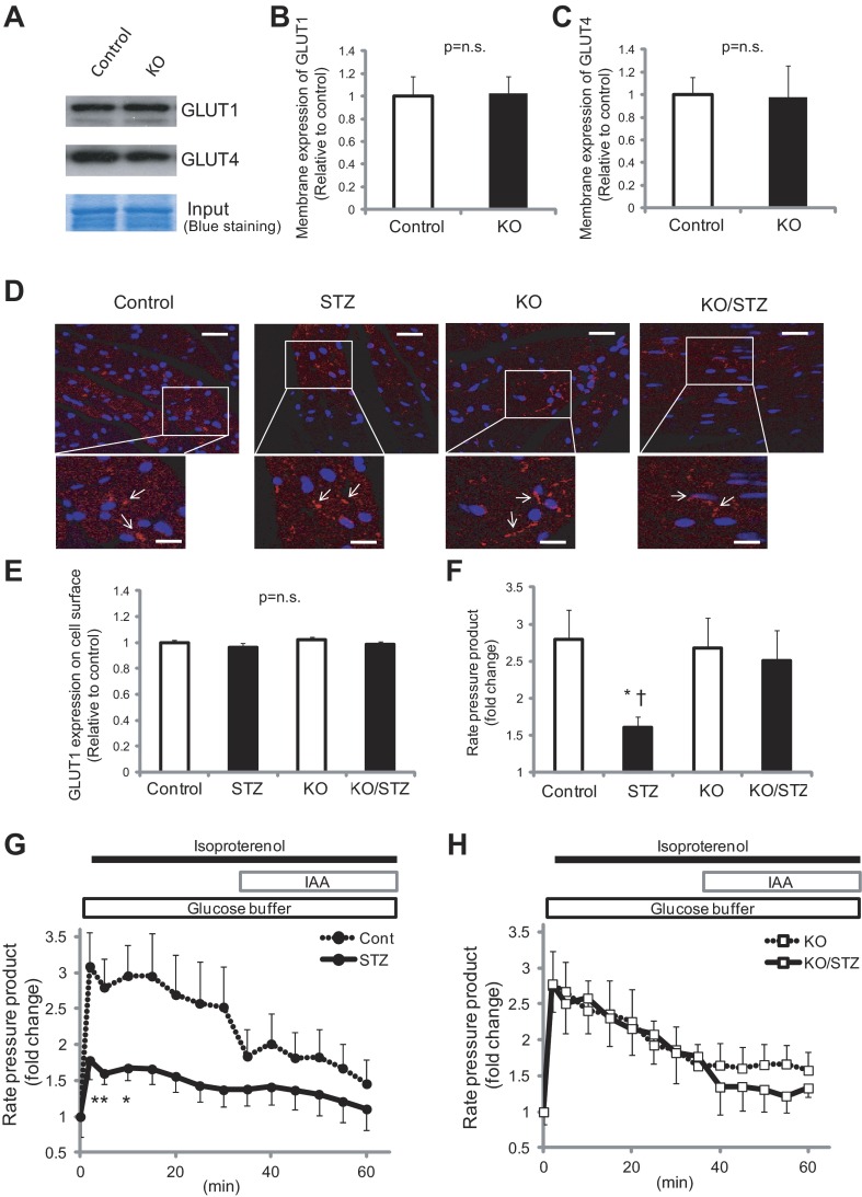
A–E: GLUT1 levels on the cell surface were comparable between Txnip-KO and wild-type myocardium in nondiabetic and streptozotocin (STZ)-induced diabetic conditions in the fed state. A: GLUT1 and GLUT4 protein expressions on the cellular membrane fraction were analyzed by Western blot. B and C: signals were quantified by densitometry of scanned autoradiographs. D: heart sections were stained with GLUT1 and secondary antibody conjugated to Alexa Fluor 594. Slides were then costained with DAPI and analyzed with confocal microscopy. Scale bars, 40 μm (lower magnification) and 10 μm (higher magnification). E: GLUT1 expression on the cell surface was quantified with ImageJ. P = N.S. among groups. F–H: functional reserve afforded by Txnip-KO hearts with diabetes is related to glucose utilization during β-adrenergic stress. Cardiac phenotype was analyzed by isolated, perfused mouse hearts with the perfusion buffer containing glucose as the sole oxidative energy substrate. Thus mechanical function was primarily supported by glucose. Changes in rate-pressure product (RPP) in response to isoproterenol (0.05 μM) were recorded in wild-type (G) or Txnip-KO (H) mice. Glycolytic inhibition with iodoacetate (IAA; 100 μM) caused a decline in mechanical function in both genotypes. The acute increase in RPP at 5 min (F) was significantly less in STZ-induced diabetic wild-type hearts (STZ) than in nondiabetic wild-type hearts (Control) or in Txnip-KO hearts without (KO) and with (KO/STZ) diabetes. Values are means ± SE. n = 3–9. *P < 0.05 vs. control (by 2-way ANOVA and unpaired t-test); †P < 0.05 vs. KO/STZ (by unpaired t-test).

