Fig. 3.
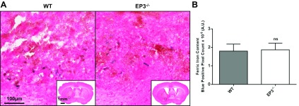
Effect of EP3 receptor deletion on brain ferric iron content following ICH. Perls' iron staining of coronal brain sections collected at 72 h post-ICH was performed to evaluate total brain ferric iron content in WT and EP3−/− mice. A: representative high magnification images showing ferric iron accumulation (blue) in perihematomal regions of WT (left) and EP3−/− (right) mice. Square selections in the inserts denote the location of magnified regions. No Perls' iron staining was observed in the contralateral hemisphere of WT or EP3−/− mice. B: blue-positive pixel count analysis showed no significant difference in total brain ferric iron content between the WT and EP3−/− groups. The comparison includes n = 11 WT and n = 13 EP3−/− mice; ns, not significant.

