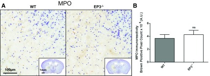
Fig. 6.
Effect of EP3 receptor deletion on peripheral neutrophil infiltration following ICH. At 72 h after ICH, immunohistochemical staining for myeloperoxidase (MPO) was used to evaluate peripheral neutrophil infiltration in WT and EP3−/− mice. A: representative high magnification images of coronal brain sections showing MPO-positive cells (brown) diffusely localized within injured brain areas. Square selections in the insets denote the location of magnified regions. No neutrophils were seen outside of the injured brain areas for any of the mice in the study. B: brown-positive pixel count analysis showed no difference in neutrophil infiltration between WT and EP3−/− mice. The comparison includes n = 11 WT and n = 13 EP3−/− mice; ns; not significant.

