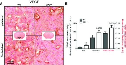
Fig. 8.
Effect of EP3 receptor deletion on VEGF expression following ICH. At 72 h after ICH, immunohistochemical staining for VEGF was used to evaluate angiogenesis/vasculogenesis in WT and EP3−/− mice. A: representative high magnification images of coronal brain sections showing the ipsilateral perihematomal region and contralateral equivalent for WT (left) and EP3−/− (right) mice. Square selections in the insets denote the location of magnified regions. B: brown-positive pixel count analysis showed significantly more ipsilateral VEGF expression in EP3−/− mice after individual signal normalization for lesion volume. A trend toward increased baseline VEGF expression, as observed by contralateral VEGF immunoreactivity, was seen in the EP3−/− mice. Ipsilateral (ipsi) and contralateral (contra) signal corresponds to left y-axis. Ipsilateral signal normalized by contralateral signal (ipsi/contra) corresponds to the right y-axis. All comparisons include n = 11 WT and n = 12 EP3−/− mice, ns, not significant; *P < 0.05.

