
Fig. 1.
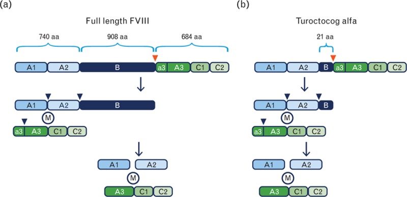
Molecular structure of FVIII and turoctocog alfa. (a) Changes to the molecular structure of FVIII from its full length (upper section) to its thrombin-activated form (lowest section). The red and blue arrowheads show the site of cleavage by the transmembrane protease furin and the three sites of thrombin cleavage, which converts FVIII to FVIIIa, respectively. M represents metal ions keeping the heavy chain and light chain together. (b) Structure of turoctocog alfa and thrombin activation. aa, amino acids; FVIII, factor VIII.