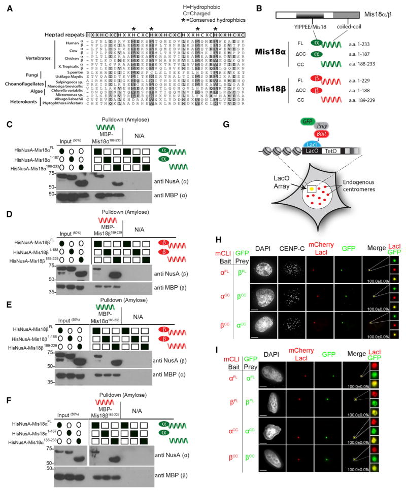
Figure 2. Mis18 Proteins Multimerize through the Conserved Coiled-Coil Domains.
(A) Diagram showing the conservation of the predicted C-terminal coiled coils. The positions of the characteristic hydrophobic (H) and charged (C) residues are shown. Shading indicates the degree of amino acid similarity (see also Figure S2A).
(B) Schematic of the Mis18 fragments used (CC, coiled coil).
(C) In vitro pull-downs using differentially tagged recombinant Mis18α fragments (Figure S1A) showing that Mis18α can homomultimerize through its predicted C-terminal coiled coil. Filled ovals or squares indicate proteins present. Proteins or affinity tags were detected by immunoblot.
(D) In vitro pull-downs of differentially tagged recombinant Mis18β fragments (Figure S1A) showing that Mis18β can homomultimerize through its predicted C-terminal coiled coil.
(E and F) In vitro pull-down using the (E) Mis18α and (F) Mis18β coiled-coil domains alone showing this domain can mediate Mis18α and Mis18β heterotypic interactions. Proteins or affinity tags were detected by immunoblot.
(G) Schematic of the LacO array system in U2OS cells. GFP-tagged prey proteins were assayed for recruitment to the noncentromeric LacO array by the mCherry LacI-fused (mCLI) bait protein.
(H) Heterotypic interactions were assayed in U2OS-LacO cells cotransfected with the indicated mCLI-Mis18α/β and GFP-Mis18α/βCC/FL constructs. Cells were stained with an anti-CENP-C antibody to mark centromeres.
(I) Homodimeric interactions were assayed in U2OS-LacO cells cotransfected with the indicated mCLI-Mis18α/β and GFP-Mis18α/βCC/FL constructs. Percentages of cells with recruitment of the prey proteins to the array are shown. Scale bars, 5 μm (see also Figures S3A–S3D and S4E).

