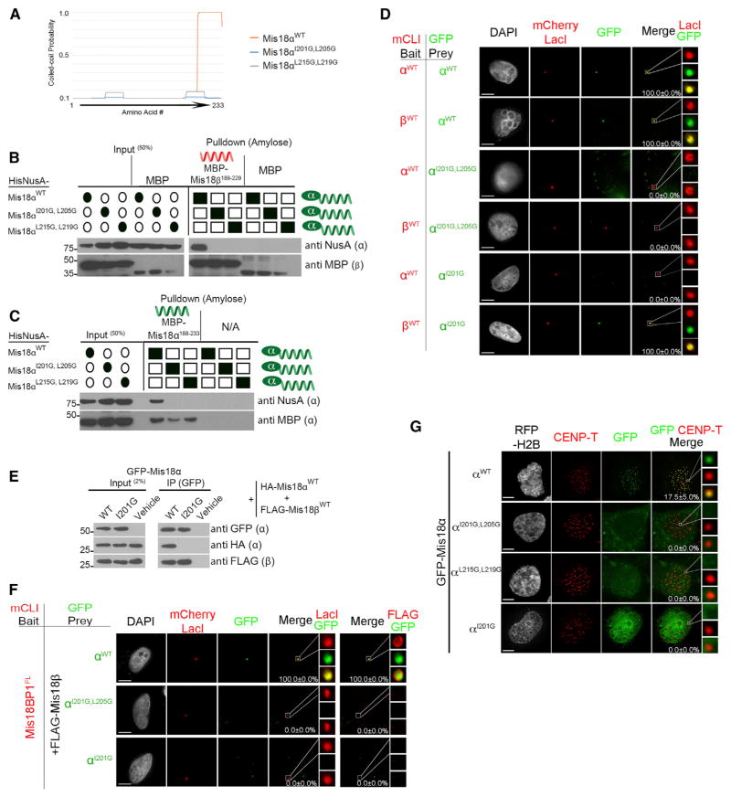
Figure 3. Mis18 Coiled Coils Are Required for Proper Centromere Localization.
(A) MARCOIL coiled-coil prediction for Mis18α with indicated mutations in the conserved hydrophobic amino acids.
(B and C) In vitro pull-down assays were conducted between recombinant (B) Mis18β or (C) Mis18α and differentially tagged Mis18α WT and coiled-coil mutants (Figure S1A). Filled ovals and squares indicate proteins present in the input and pull-downs. Proteins or affinity tags were detected by immunoblot.
(D) U2OS-LacO cells were cotransfected with mCLI-Mis18α/β and GFP-Mis18αWT/I201G,L205G/I201G or Mis18αI201G. Percentages of cells with recruitment of the prey proteins to the array are shown. Scale bars, 5 μm.
(E) Anti-GFP immunoprecipitation conducted from U2OS cells transfected with GFP-Mis18α (WT or Mis18αI205G mutant), HA-Mis18α (WT), and FLAG-Mis18β (WT).
(F) U2OS-LacO cells were cotransfected with mCLI-Mis18BP1, GFP-Mis18αWT/I201G,L205G/I201G or Mis18αI201G, and FLAG-Mis18βWT. Percentages of cells with recruitment of the prey proteins to the array are shown. Scale bars, 5 μm.
(G) U2OS cells were cotransfected with GFP-Mis18α WT or mutant constructs. RFP-histone H2B was used as a transfection marker. Centromeres were identified using anti-CENP-T antibody. The percentage of cells with GFP-Mis18 recruited to centromeres is shown ± SD. Scale bars, 5 μm (see also Figures S4A–S4D and S3E).

