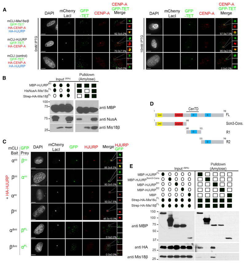
Figure 4. The Mis18α/β Complex Promotes CENP-A Deposition through a Direct Physical Interaction with HJURP.
(A) U2OS-LacO cells were cotransfected with the indicated mCLI, GFP-TET, HA-CENP-A, and HA-HJURP constructs with/without 10 mM IPTG treatment to disrupt the LacI interaction with the LacO array. Cells were stained with anti-CENP-A antibody. The LacO array was marked by GFP-TET. Recruitment of CENP-A to the LacO array can be seen in the magnified boxed regions to the right. Percentages of cells with CENP-A recruitment to the array are shown. Scale bars, 5 μm.
(B) In vitro pull-down of recombinant HJURP and differentially tagged Mis18α and Mis18β. Filled ovals and squares indicate proteins present in the input and pull-downs. Proteins or affinity tags were detected by immunoblot.
(C) U2OS-LacO cells were cotransfected with mCLI-Mis18α/βCC, GFP-Mis18α/βCC, and HA-HJURP. Cells were stained with an anti-HA antibody. Percentages of cells with recruitment of the prey proteins to the array are shown. Scale bars, 5 μm (see also Figure S5A).
(D) Schematic of the domain structure of HJURP and the recombinant fragments used in this experiment. CenTD, centromere-targeting domain.
(E) In vitro pull-down between recombinant HJURP fragments, Mis18α, and Mis18β (Figure S1A). Proteins or affinity tags were detected by immunoblot.

