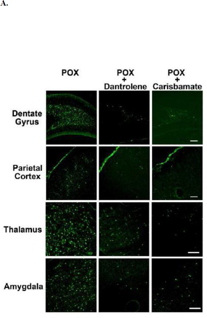
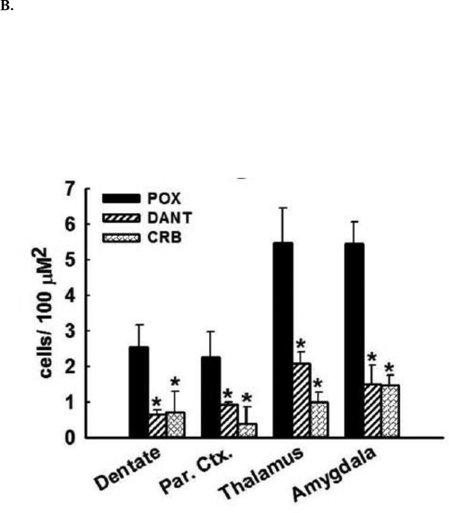
Figure 2. Neuroprotective effects of dantrolene and carisbamate following POX SE.
A. Representative photomicrographs of Fluoro-Jade C (FJC) staining in the dentate gyrus-hilus region, parietal cortex, amygdala, and thalamus of a POX rat 2 days after POX SE, and POX + dantrolene, and + carisbamate treated rats. Scale bars, 200 µm.
B. Quantitative analyses of FJC labeling. Control rats did not exhibit any FJC labeling. FJC positive cells indicative of neuronal injury were observed in hilus, amygdala, thalamus and cortex of POX rats 48-h after SE termination. Rats treated with dantrolene or carisbamate showed significantly less FJC labeling in these brain regions (*p<0.05 compared to control, t-test, n= 6 rats).


