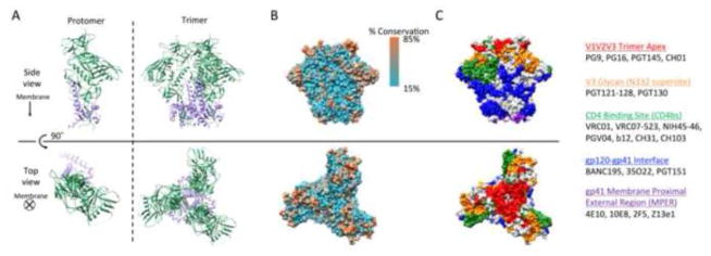
Figure 1.
Structure of the HIV Envelope glycoprotein based on the crystal structure of BG505 SOSIP.664 (PDB ID: 4TVP). A] Ribbon representation of HIV gp140 protomer and trimers composed of gp120 (green) and gp41 (purple) subunits. B] Surface structure of HIV Env trimer colored by degree of conservation in the 2014 HIV Sequence Compendium from the LANL database [331]. C] Surface epitope domains colored by contact residues and residues ≤ 4Å from contact residues: V1V2V3 loop/trimer apex (red); V3 glycan-N332 supersite (orange); CD4 binding site (green); gp120-gp41 interface (blue); gp41 membrane-proximal external region (purple). Common bNAbs against each epitope domain are listed. Glycans are not shown, but importantly mask a significant portion of the viral Env surface. Images were generated using the UCSF Chimera package.[332]

