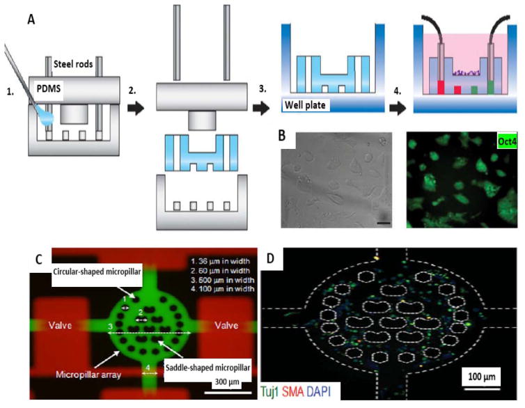
Figure 3.
Fabrication of microchips and ESC culture. A) Schematic illustration of fabrication of the hydrogel microfluidic chip. 1. Mold fabrication and injection of hydrogel monomers. 2. Gelation, and mold removal. 3. Transfer to a well plate. 4. Cell seeding and system activation for cell-based assays. B) ESC culture on the surface of the chip (reproduced from ref. 113. Copyright 2015 with permission from nature publishing group). (C) Dual-micropillar-based microfluidic platform containing microvalves. (D) Neural-like cells in microchip with stained Tuj1 (antineuronal classIII, β_tubulin), SMA (anti-α-smooth muscle actin) and DAPI (blue-fluorescent DNA stain) (reproduced from ref. 95. Copyright 2015 with permission from John Wiley & Sons.).

