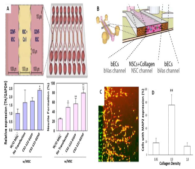
Figure 5.
Investigation of NSCs biology via microfluidics. A) Microfluidic platform for in vivo paracrine signaling mimicking for enhanced NSC differentiation. Top: Schematic illustration of Microfluidic array for NSCs and GDNF-MSCs coculturing. Bottom (left): qRTPCR experiments with upregulated expression of the TH marker in hNSCs. Bottom (right): Neurite formation level of GDNF-hMSCs in comparison with control groups (Reproduced from Ref. 108. Copyright 2015 with permission from Elsevier). B) Schematic illustration of microdevice for interaction of cultured NSCs with bVas (Reprinted from Ref. 115. Copyright 2015 with permission from John Wiley & Sons). C and D) Neural stem/progenitor cells migration and differentiation in the collagen matrix. C) Fluorescent labeling (red) of MAP2 protein expression in appropriate collagen density (0.9 mg/ml). D) Neural stem/progenitor cells percentage with MAP2 expression within different collagen matrices (Reproduced from Ref. 128. Copyright 2015 with permission from the Royal Society of Chemistry).

