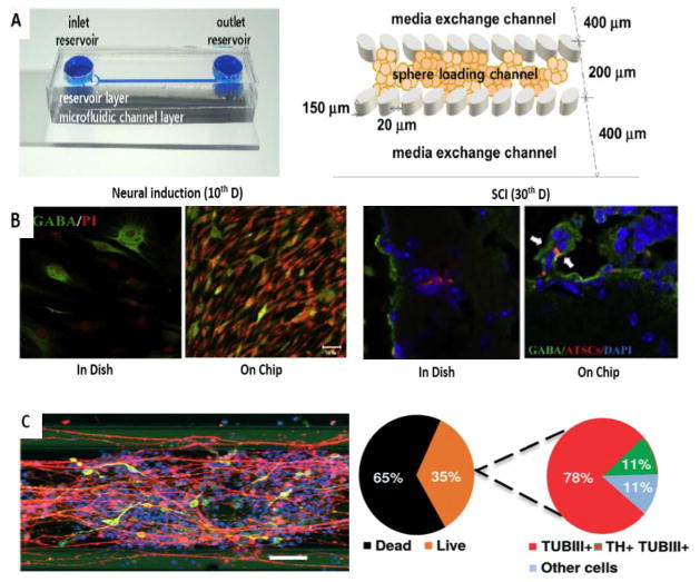
Figure 6.
Application of microfluidics for other types of stem cells. A–B) Neuronal differentiation in a gel-free microfluidic chip. A) Microchip with two inlets (exchanging medium and loading neurospheres) and one outlet (left) and schematic concept of immobilization of the neurospheres in the sphere-loading channel with elliptical pillars (right). C) Efficient neurogenesis of hATSC neurospheres into GABA secreting neurons (60% of DAPI) in comparison to dish culture (30% of DAPI) (left). High rate of neurogenesis to give motor neurons (NF160+) in chip culture of hATSC neurospheres compared to dish culture in the lesions of mouse injured spinal cord tissue (10–15% transdifferentiation of engrafted cells in comparison with less than 5%, respectively) (right) (Reproduced from Ref. 111. Copyright 2015 with permission from Elsevier). C) Immunostaining of differentiated neurons with nuclei, TUBβIII and TH stains in a microfluidic bioreactor for differentiation of human NEPSCs into neurons (left). Scale bar: 100μm. Live–dead cell correlation and efficiency of differentiation (right). (Reproduced from Ref. 58. Copyright 2015 with permission from the Royal Society of Chemistry).

