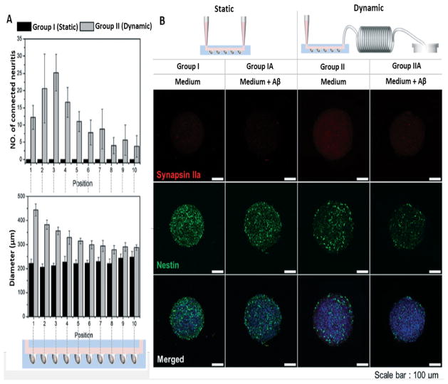
Figure 7.
Neural network formation and neurotoxic effects of amyloid-β. A) Average number of neurites extending (upper graph) and average size of neurospheroids (lower graph) in different microdevice sections of two main group. B) Destruction of neural networks shown by immunofluorescence images staining synapsin IIa (synaptic marker) and nestin (neural progenitor/stem cell marker) in neurospheroids in different groups (Scale bar:100 μm). (Reproduced from Ref. 147. Copyright 2015 with permission from the Royal Society of Chemistry).

