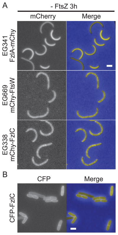
Figure 1. FzlC localizes to membranes in C. crescentus and E. coli cells.
(A) Fluorescence and merged micrographs of cells depleted of FtsZ for 3 h and expressing mCherry fusions to the indicated proteins induced with vanillate for 2 h. FzlA is diffuse in the cytoplasm (top row) while FtsW and FzlC display a patchy peripheral localization typical of membrane-associated proteins (middle and bottom rows). (B) Fluorescence and merged micrographs of cells producing CFP-FzlC after 2 h induction with 1% L-arabinose in E. coli. CFP-FzlC localizes to the periphery, indicating membrane association. Scale bars = 2 μm.

