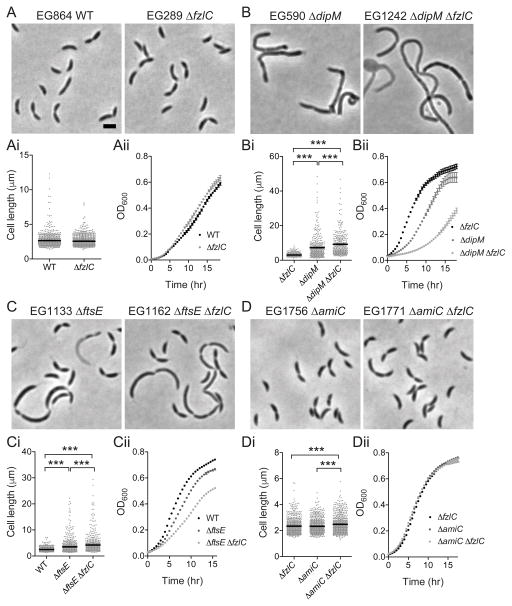
Figure 7. Deletion of fzlC has synthetic interactions with non-essential division genes dipM, ftsE, and amiC.
(A–D) Phase contrast micrographs of cells with or without fzlC in WT, ΔdipM, ΔftsE, or ΔamiC backgrounds. (Ai-Di) Cell length of strains in (A–D) (see Table S3 for sample sizes). Error bars represent the mean cell length ± SEM, *** = p < 0.001, one-way ANOVA. (Aii-Dii) Growth curves of strains shown in (A–D). Scale bars = 2 μm.

