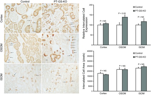
Fig. 3.
GS expression in intercalated cells in the cortex, OSOM, and inner stripe of the outer medulla (ISOM). Intercalated cell GS immunolabel (arrows) was present in PT-GS-KO mice in a pattern similar to that observed in control mice (left). In the OSOM, intercalated cell GS immunolabel intensity appeared greater in PT-GS-KO mice than in control mice. Quantitative immunohistochemistry (top right) confirmed this observation. There was no significant difference in intercalated cell GS immunolabel intensity in either the cortex or ISOM. PT-GS-KO did not significantly alter intercalated cell mean area in either the cortex, OSOM, or ISOM (bottom right). n = 4 mice/genotype. NS, not significant.

