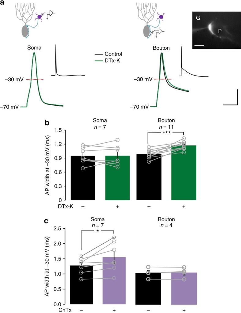
Figure 1. Compartment-specific roles of K+ channels in action potential shape in cerebellar basket cells.
(a) Representative action potentials recorded from the soma (left) or bouton (right) of cerebellar basket cells, before (black trace) and after (green trace) Kv1.1 blockade with DTx-K. Top: schematics illustrating recording sites. Inset: action potentials at slow time base, showing a prominent ADP at the bouton. Scale bar, 20 mV; 2 ms (main traces); 40 mV, 200 ms (insets). The fluorescence image at right shows a cerebellar basket cell bouton labelled with Alexa 568. G and P indicate granule cell and Purkinje cell layer, respectively. Scale bar, 10 μm. (b) Summary data showing selective effect of DTx-K on presynaptic spike width measured at –30 mV. Circles show individual experiments. (c) BKCa blockade with ChTx led to somatic action potential broadening. *P<0.05, ***P<0.001; paired t-tests.

