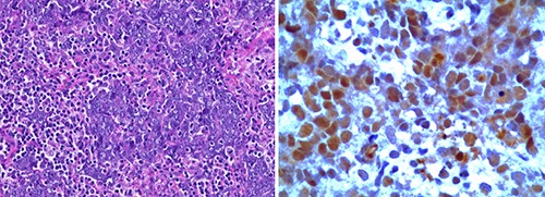
Figure 2.

Diagnosis of nuclear protein of the testis (NUT) midline carcinoma. A) Undifferentiated small cells with foci of squamous differentiation (Hematoxylin & Eosin, 20×). B) Immunohistochemistry of tumor cell nuclei showing speckled staining for NUT (250×) using anti-NUT rabbit polyclonal antibody (clone C52, 1:100).
