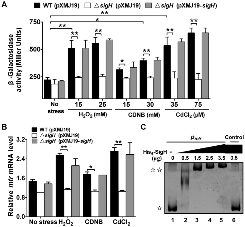
Figure 8. Positive regulation of C. glutamicum mtr expression by SigH.
(A) β-Galactosidase analysis of the mtr promoter activity was performed using the transcriptional Pmtr::lacZ chromosomal fusion reporter expressed in the wild-type, ΔsigH mutant, and complemented strain ΔsigH(pXMJ19-sigH). Exponentially growing C. glutamicum cells (100 μl) induced with different toxic agents at the indicated concentrations for 30 min were added to the enzyme reaction system. β-Galactosidase activity was assayed as described in “Methods.” Mean values with standard deviations (error bars) from at least three repeats are shown. *P ≤ 0.05. **P ≤ 0.01. (B) qRT-PCR revealed that expression of mtr was under strict positive regulation by SigH. Exponentially growing C. glutamicum cells were exposed to different toxic agents at the indicated concentrations for 30 min. Levels of mtr expression were determined by qRT-PCR. The mRNA levels are presented relative to the value obtained from wild-type cells without treatment. Mean values with standard deviations (error bars) from at least three repeats are shown. *P ≤ 0.05. **P ≤ 0.01. (C) Interactions between SigH and the mtr promoter were analysed by EMSA. Increasing amounts of SigH used were 0, 0.5, 1.5, 3.0, and 3.5 μg. As a negative control, a 400-bp fragment from the mtr coding region amplified with primers Control-F and Control-R, replacing the 400-bp mtr promoter, was incubated with 3.5 μg of His6-SigH in the binding assay. (☆) free DNA and (☆☆) major DNA-protein complex.

