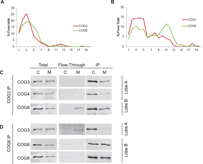
Figure 1. Membrane associated COG subunits are arranged as octamers and tetramers.
HeLa cell lysates were separated into cytosol [C] and membrane [M] fractions by differential centrifugation. Efficient separations were confirmed by SDS-PAGE using antibodies against GAPDH (cytosolic enzyme) and Vti1a (transmembrane protein) (Supplemental Figure 1C). HeLa cytosol (A) or Triton-X100 soluble membranes (B) were loaded onto a Superose 6 10/300 GL and 0.5 mL fractions were collected. Fractions were concentrated by TCA precipitation, separated on a SDS-PAGE gel, transferred to a nitrocellulose membrane, and probed with anti-COG3 and anti-COG8 antibodies. Soluble COG subunits peaked in early fractions (fractions 2–4; Ve = ~9–10.5 mL) indicative of the size of the COG complex octamer. Membrane COG subunits peaked in fractions 2–4 (Ve = ~9–10.5 mL) and also in fractions 10–12 (Ve: ~12.5–14 mL) corresponding in size to a tetramer. HeLa cytosol and membrane fractions were immunoprecipitated (IP) with anti-COG3 (C) and anti-COG6 antibodies (D). Immunodepleted/flow-through (F/T) and IP fractions were separated by SDS-PAGE and blotted with antibodies to COG3, COG4, COG6, and COG8. IP of lobe A subunit COG3 immunodepleted lobe B subunit COG8 from cytosol, but not membrane fraction. IP of lobe B subunit immunodepleted lobe A subunits COG3 from cytosol, but not membrane fraction.

