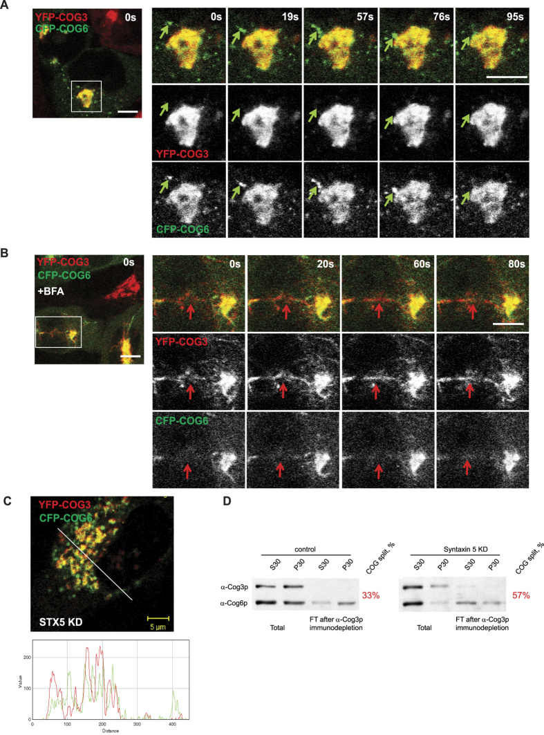
Figure 2. COG sub-complex lobe B localizes to Golgi and vesicle membranes.
(A) Static live cell images of HeLa cells stably expressing lobe A subunit YFP-COG3 (red) and lobe B subunit CFP-COG6 (green). CFP-COG6 partially localizes to smaller vesicle-like structures separate from YFP-COG3 and Golgi apparatus (A). Green arrows indicate structure labeled with CFP-COG6 but not YFP-COG3 moving towards and fusing with the Golgi. (B) Static live images of HeLa YFP-COG3/CFP-COG6 cells treated with 0.25 μg/l BFA for 15 min. Red arrow indicates a YFP-COG3 labeled tubule which does not contain CFP-COG6. Bar, 10 μm. (C) HeLa YFP-COG3/CFP-COG6 cells depleted for Golgi t-SNARE STX5 were fixed and analyzed by IF. Line plot for overlap between red and green channels is shown measuring the relative value of signal intensity (y-axis) over the distance measured in pixels (x-axis). Bar, 5 μm. (D) HeLa control and STX5 depleted cells were separated by centrifugation on S30 and P30 fractions and immunodepleted using anti-COG3 antibodies. Initial and immunodepleted fractions were separated by SDS-PAGE and blotted with antibodies against COG3 and COG6. Numbers indicate percentage of lobe B COG6 protein not associated with lobe A COG3.

