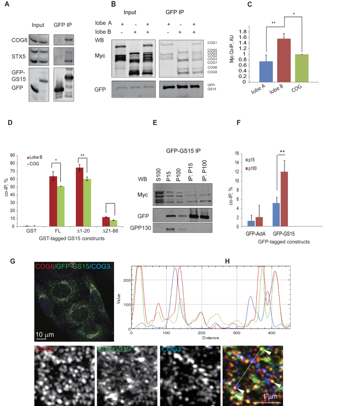
Figure 4. COG lobe B sub-complex preferentially interacts with GS15.
(A) GFP-GS15 interacts with endogenous COG8. HEK293T cells were transiently transfected with GFP-GS15 or GFP then immunoprecipitated. IPs were probed by western blot with antibodies against COG8, STX5, and GFP. (B) GFP-GS15 preferentially interacts with lobe B in vivo. HEK293T cells were transiently transfected with COG-Myc multi-expression plasmids (lobe A expression, lobe B expression, or the whole COG complex). 24 h after COG-myc transfection cells were transfected with GFP-GS15. 24 h post transfection cellular lysates were immunoprecipitated. IPs were probed by western blot with antibodies against Myc and GFP. (C) Co-IP efficiency values were generated by calculating the relative density of the co-IP Myc signal relative to the density of the input Myc signal and the recovered GFP signal in four independent experiments. (D) COG-GS15 in vitro binding assay. Glutathione beads were incubated with recombinant GST, GST-tagged full-length (FL) GS15 cytoplasmic domain (aa 1–86), GS15 with deletion of N-terminal domain (Δ1–20), and deletion of SNARE domain (Δ21–86). Bound proteins were eluted from beads, and probed by western blot with Myc antibodies. IP efficiency values were generated by normalizing the density of the IP Myc signal to the density of the whole COG/Lobe B input Myc signal. (E) Lobe B interacts with GS15 on vesicle membranes. HEK293T cells were transiently transfected with lobe B and GFP-GS15. 48 h after transfection, cells were collected and fractionated into large membrane (P15), small membrane (P100), and cytosol (S100) by differential centrifugation. The membrane pellets were solubilized with Triton-X100 and then incubated with GBP-beads. IPs were probed by western blot with anti-Myc, anti-GFP, and anti-GPP130. (F) Co-IP efficiency values were calculated by dividing co-IP by input in four independent experiments. (G) HeLa cells stably expressing GFP-GS15 were stained for COG3 and COG8 and imaged using the Zeiss ELYRA S1. Line plots are shown measuring the relative value of signal intensity (y-axis) over the distance measured in pixels (x-axis). White arrows in the merged image indicate GS15 and COG8 co-localizing on vesicle-like structures. Bar, 10 μm (inset 1 μm).

