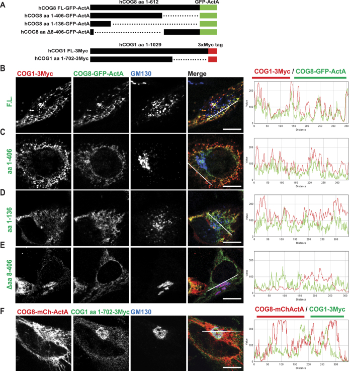
Figure 6. The C terminus of COG1 interacts with the N terminus of COG8.
(A) Schematic of the constructs used in the mitochondrial relocalization assay: Full length (FL) COG8, COG8 aa 1–406, COG8 aa 1–136, or COG8 Δaa8–406 tagged with GFP-ActA. COG1 full length (FL) or 1–702 tagged with 3Myc. HeLa cells were transiently transfected with plasmids as follows: (B) hCOG1-Myc and hCOG8-GFP-ActA, (C) hCOG1-Myc and hCOG8(1–406)-GFP-ActA, (D) hCOG1-Myc and hCOG8(1–136)-GFP-ActA, (E) hCOG1-Myc and hCOG8(Δ8–406)-GFP-ActA, (F) hCOG8-mChActA and hCOG1(1–702). 24 h after transfection cells were fixed, and then stained with antibodies to Myc and GM130 and analyzed by confocal microscopy. Line plots for overlap between red and green channels are shown. Size bar, 10 μm.

