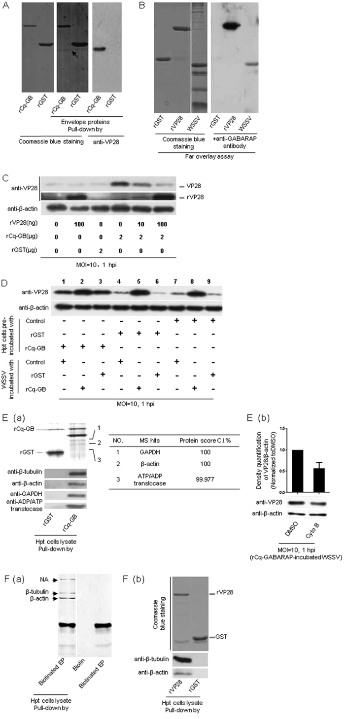
Figure 6. Protein interactions between rCq-GABARAP, WSSV and Hpt cell led to viral entry promotion.

(A) rCq-GABARAP bound to the viral envelope protein VP28 in pull-down assays. The pull-down samples were resolved by SDS-PAGE followed by Coomassie blue staining (middle panel) and immunoblotting with an anti-VP28 monoclonal antibody (right panel). The arrow indicates the viral envelope protein VP28 which was further determined by MALDI TOF/TOF mass spectrometry. (B) rCq-GABARAP binding to the viral envelope protein VP28 confirmed by a far-western overlay blotting assay. The protein samples in the SDS-PAGE gel (left panel) were transferred to a PVDF membrane, which was then incubated with rCq-GABARAP and detected by immunoblotting with an anti-GABARAP antibody (right panel). (C) rCq-GABARAP-promoted WSSV entry attenuated by incubation with rVP28. (D) Synergistic effect on enhanced WSSV entry by rCq-GABARAP via targeting both WSSV and Hpt cell. (E) Interactions of rCq-GABARAP with the cytoskeleton components of Hpt cell. (a) The proteins pulled down from the Hpt cell lysate by rCq-GABARAP as detected by silver staining or Western blotting. (b) Suppression of rCq-GABARAP-mediated WSSV entry via pre-treatment of the cells with cytochalasin B as determined by immunoblotting to quantify the viral envelope protein VP28 (low panel). The band intensities of VP28 and β-actin from three independent experiments were analyzed using the Quantity One program (upper panel). (F) Interactions of WSSV with Hpt cell. (a) Binding of Hpt cell proteins to biotinylated WSSV envelope proteins (EP) was examined by pull down assay. The specific protein bands that only bound to the biotinylated envelope proteins indicated with arrows were selected for mass spectroscopy determination. NA, data not available. (b) Binding of β-tubulin or β-actin with rVP28. Hpt cell lysate was prepared and used for pull-down assays with rVP28 or GST as a control. The sample was analyzed using an anti-β-tubulin or anti-β-actin antibody. Note: The results in B and F(a) were assembled by rearranging and joining images of different lanes that originated from the same film. All results were observed in at least three independent experiments, and one set of representative results is shown.
