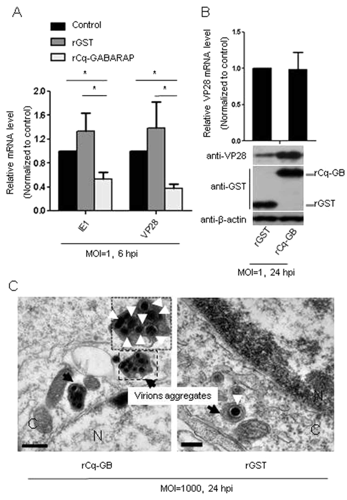
Figure 7. Suppression of WSSV replication by rCq-GABARAP.

WSSV at different MOIs was incubated with rCq-GABARAP as indicated and then inoculated into Hpt cell cultures. (A) Quantification of WSSV transcripts (IE1 and VP28) by qRT-PCR. The Hpt cells were infected with virus pre-incubated with rCq-GABARAP or rGST, and the relative gene transcript expression of IE1 and VP28 was compared to that obtained with virus without incubation with the recombinant protein. (B) WSSV entry but not viral replication (upper panel) enhanced by rCq-GABARAP at a late stage of viral infection (24 h) (lower panel). Increased VP28 protein levels were detected in Hpt cell infected with WSSV pre-incubated with rCq-GABARAP as determined by immunoblotting against the viral envelope protein VP28 at 24 hpi. (C) An increased number of WSSV virion aggregates was found in the Hpt cell infected with WSSV that had been pre-incubated with rCq-GABARAP compared to those infected with the rGST pre-incubated virus as visualized by TEM. Bars: 0.5 μm in the left image, 0.2 μm in the right image; C, cytoplasm; N, nucleus. WSSV virions are indicated with white arrowheads. WSSV virion aggregates are indicated with black arrows. All of the results were observed in at least three independent experiments, and one set of representative results is shown. The data are presented as the mean ± SEM from at least three independent experiments and were analyzed by Student’s t test (*P < 0.05).
